Myosin V
Structure
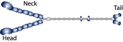
Myosin V is a multimeric protein that consists of 16 polypeptide chains. The overall structure can be divided into 3 major subdivisions the: head, neck and tail (Cheney et al., 1993).

The head: The myosin V head domain, sometimes referred to as the motor domain is ~80 kD and contains both the actin binding and ATP hydrolytic activities. Sequence comparison of the motor domains from members of the myosin super-family reveal 3 primary regions of variability (Cope et al., 1996), which have been proposed to be related to the functional and enzymatic differences between myosins.
The neck: Many members of the myosin superfamily have a neck domain that contains tandem copys of the calmodulin-binding consensus sequence (IQXXXRGXXXR). Variations on the sequence and number of motifs are found among all myosins (Mermall et al., 1998). Myosin V represents the giraffe of myosins with a neck that contains 6 IQ motifs, each of which has the potential to bind either calmodulin or an essential light chain (ELC).
The Tail: The tail region of myosin V consists of regions of alpha helical coiled coil that are interrupted by globular regions. The coiled coil regions are thought to play a role in dimerization. The function of the globular regions is not known but they are believed to be involved in cargo binding.
Function
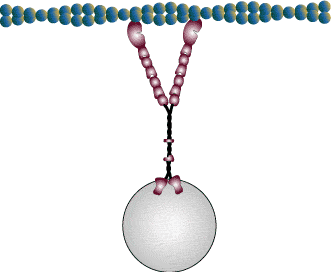
The phenotype of myosin V mutations in yeast and mice suggest that myosin V may function in organelle transport.
The mouse dilute locus encodes myosin V and several mutations have been identified that have varying degrees of pigmentary dilution and nervous system defects. (Jenkins et al., 1981). The pigmentary dilution results from disruption of melanosome transport and the nervous seizures are thought to result from disrupted transport of smooth endoplasmic reticulum into the dendritic spines of purkinje cells.
We used single molecule (Molecular Motors Group, Warshaw Lab) and ensemble motility (Molecular Motors Group, Warshaw Lab)experiments to study the unique properties of myosin V that allow it to perform its role as an organelle transporter.
Above figures modified from the Myosin Home Page.